Services
SERVICES
SOLUTIONS
TECHNOLOGIES
Industries
Insights
TRENDING TOPICS
INDUSTRY-RELATED TOPICS
OUR EXPERTS

April 3, 2025
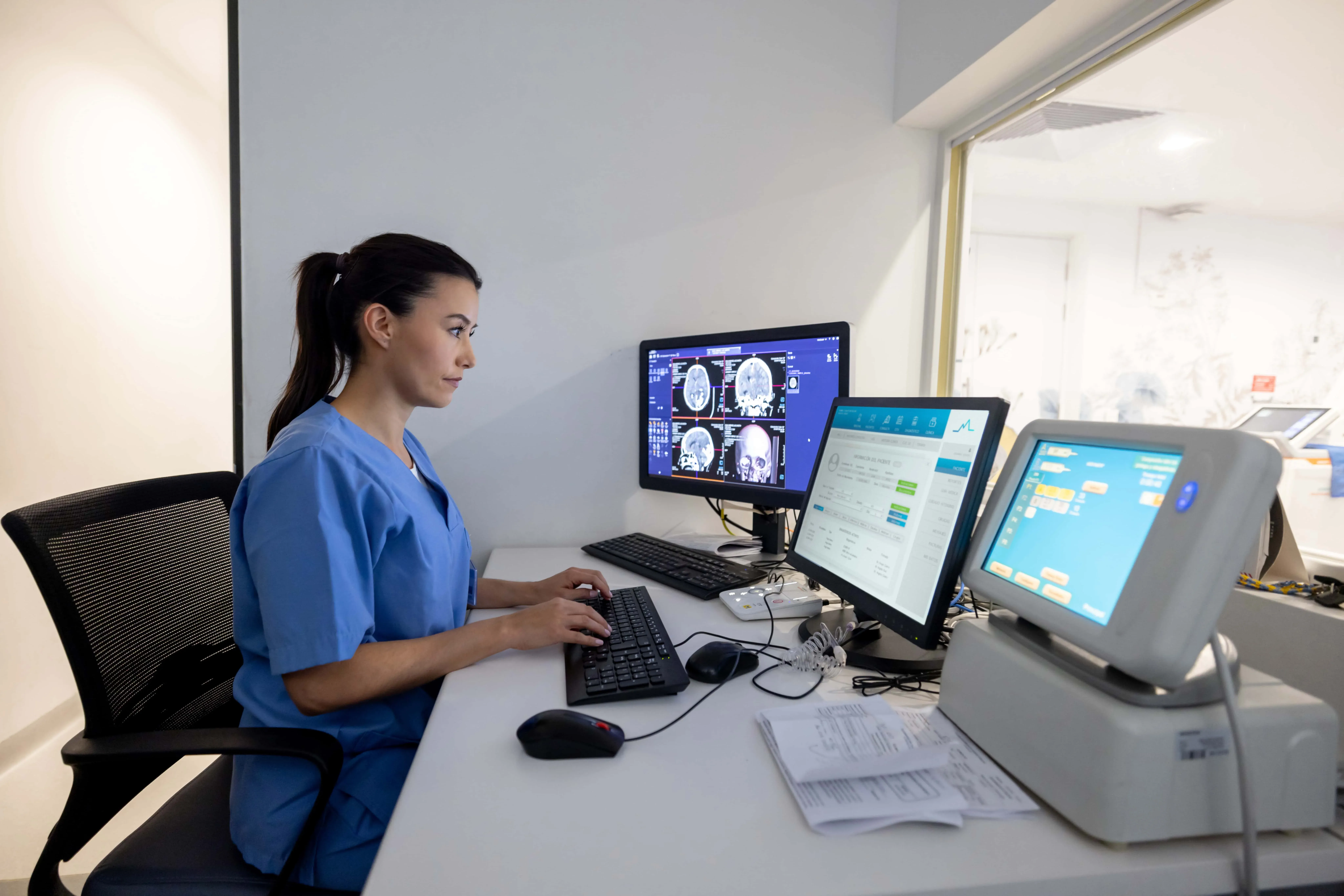
Medical image analysis software can assist healthcare companies by performing the following tasks:

Discerns important details in images by reducing noise, adjusting contrast, and removing artifacts and blurs to improve the quality of medical images and make them more suitable for analysis.
Determines whether objects of a specific class, such as tumors, are present in the studied area and then localizes their exact coordinates, including localizing them in a 3D model based on 2D images.
Recognizes contours of particular organs or anatomical structures in an image and discovers any anomalies in organ structures such as traumas and lesions.
Studies the correspondence among multiple images and tracks changes in patients’ lesions by combining data collected from diverse sources and at different points in time into a single dataset.
Separates medical images that contain anomalies from the ones that don’t to detect different types of cancer and other diseases.
Measures various parameters, such as the size, volume, texture, shape, density, or intensity of biological structures, to identify abnormalities in an image and monitor disease progression.
Provides clinicians with immersive visualization and detailed analysis of complex anatomical features by combining multiple 2D images into one single image, dividing 3D or 4D reconstructions into 2D components, and segmenting 2D images.
AI algorithms are powered by various technologies that enable real-time pattern recognition, workflow automation, and big data analysis. The common AI technologies used in biomedical image analysis include:
Deep learning frameworks |
|
|
| |
|---|---|---|---|---|
Computer vision technologies |
|
|
|
|
Cloud providers |
|
|
|
|
Medical image analysis software reveals hidden patterns and enables clinicians to make more accurate diagnoses as well as predict serious and terminal diseases years before they manifest, generating personalized recommendations and treatment plans.
Thanks to the automated capture and analysis of multi-modal images and the enhanced ability to identify subtle patterns and abnormalities, healthcare professionals can study images more quickly and make fewer mistakes.
Sophisticated machine learning algorithms detect abnormality in human cells, tissues, and organs and analyze genetic information. They ascertain an individual’s susceptibility to particular diseases like cancer, allowing doctors to determine and address care gaps and improve health metrics.
By facilitating cross-patient comparisons and post-factum analysis, medical image analysis software helps research institutions accelerate study processes and uncover new disease patterns and biomarkers to develop better treatments and improve public health.
RapidAI is an AI-powered software platform designed to simplify the examination, processing, and analysis of CT scans, including non-contrast CT, CT angiography, and CT perfusion images. Being the leader in brain imaging analysis, RapidAI incorporates a number of static AI-derived algorithms, such as Rapid ICH, Rapid ASPECTS, Rapid CTA, Rapid LVO, Rapid CTP, and Rapid HVS. It features solutions for perfusion imaging, cerebral aneurysm triage and management, pulmonary embolism detection, and report generation, helping doctors triage patients and determine the best course of treatment. The system can be accessed from smartphones and desktops, allows care teams to communicate securely, and automatically generates reports and notifications of suspected health anomalies.
It’s an intelligent care coordination solution that enables quick and precise disease detection, using AI to read imaging (CT scans, EKGs, echocardiograms, etc.) and other patient data and automatically sharing relevant scans and data between care teams. It’s an FDA-approved software that covers 12 disease areas, including neurology, cardiology, and vascular domains, with oncology being a new focal point.
Viz.ai works on 13 AI care coordination algorithms, sending AI alerts of suspected diseases, supporting AI-powered screening and 3D imaging, and facilitating real-time care team collaboration. To expand product capabilities, the company established a partnership with Microsoft in 2024 to integrate its AI models and care coordination solution with Precision Imaging Network, a component of Microsoft Cloud for Healthcare.
Aidoc offers a suite of AI-enabled, FDA-cleared products that assist clinicians with diagnosing patients and conducting full-body CT scans. The platform analyzes medical images of the head, neck, chest, and abdomen and is capable of identifying difficult-to-detect abnormalities that require immediate attention, including large-vessel occlusions (LVOs), intracranial hemorrhage and pulmonary embolism.
In 2024, the company also launched new AI solutions for assessing cardiovascular and neurological conditions, notifying and triaging urgent pathologies like vessel occlusion, aortic dissection, vertebral compression fractures, and malpositioned endotracheal tubes. In total, Aidoc’s aiOS™ platform now includes 26 AI solutions deployed via the Aidoc aiOS™ platform.
The AGFA HealthCare Enterprise Imaging Platform features advanced, native diagnostic imaging features combined with intelligent workflow orchestration capabilities to help diagnosticians interpret images across radiology, cardiology, pathology, and other disciplines. It can be seamlessly integrated with EHRs and PACS to create a comprehensive Imaging Health Record™ (IHR).
Agfa HealthCare offers a fully managed SaaS solution as well as supports customer-managed cloud deployment. The platform provides advanced visualizations, automated triage, smart hanging protocols, and automated reporting and notification of critical findings, all powered by AI. It also comes with business intelligence tools to gather, organize, analyze, and manage clinical, financial, and administrative data.
Plus, AGFA HealthCare’s Streaming Client solution provides a unified environment that supports real-time chat and screen sharing with radiologists. Agfa also offers modular solutions like VNA (Vendor-Neutral Archive) and cross-enterprise worklists to unify imaging data across multiple locations.
Imaging Insights is a tool provided by GE HealthCare that creates the consolidated views of data from MR and CT systems to help radiologists access and analyze imaging data. Imaging Insights, which is based on GE HealthCare’s Applied Intelligence platform, combines data from the machines and the Radiology Information System (RIS) to provide actionable insights that improve healthcare workflows, boost productivity, and help with decision-making.
The software simplifies protocol standardization and dose management and helps identify variations in practices and staff performance for healthcare providers to create relevant training programs as well as spot variations in equipment usage for better resource coordination.
When implementing image analysis software, healthcare organizations should be ready to tackle the following issues.
Challenge | Possible solution | |
Dataset bias | Medical imaging datasets used to train the model can contain erroneous or insufficient information about
underrepresented groups or patients with unusual medical diseases, leading to incorrect interpretation,
model bias, and inappropriate treatments. | Gather information from diverse demographic groups, regions, and contexts. Create more representative training datasets with data augmentation techniques or data filtering, specifically adding underrepresented examples to the data or removing undesirable samples. Establish rigorous data collection, processing, and review processes. |
|---|---|---|
Security & compliance challenges | Medical image analysis software processes enormous volumes of sensitive patient information, which is
hard to keep confidential, raising the possibility of data breaches or unwanted data access. | Establish robust data security measures, such as data anonymization, encryption, and access restrictions, and choose software that complies with relevant data privacy laws, such as HIPAA or GDPR. To reduce ethical concerns, obtain informed consent from patients when collecting data. |
Pushback from healthcare professionals | Some radiologists can hesitate to use medical image analysis software due to its complexity and
additional efforts involved in maintaining data correctness and acquiring patient consent to collect data. | Conduct training sessions and workshops, demonstrating successful use cases of medical image analysis software and educating medical personnel on the specifics and usage of these tools. |
Lack of data interpretability | Failure to understand the reasoning behind a deep learning model’s decision-making process lowers trust
in the algorithm’s prediction, hinders clinical acceptance, and complicates the identification and
mitigation of biases within the data and model. | Use visualization approaches and heatmaps to show which areas of an image drive the model judgments, explainable AI (XAI) methods, and attention mechanisms to draw the doctor’s attention to key aspects of an image and explain the model’s decision-making process. |
| By 2029, the global market for diagnostic imaging devices is expected to generate $59.25 billion in revenue | |
|---|---|
| With a compound annual growth rate (CAGR) of 7.8%, the global medical image analysis software market is projected to reach $4.5 billion by 2027 | |
| The top 5 countries by revenue generated in the diagnostic imaging devices market in 2029 will be the USA ($14,330 million), Germany ($4,935 million), Japan ($4,388 million), China ($4,286 million), and the United Kingdom ($2,997 million) | |
| As much as 80% of vital healthcare data remains unstructured in the form of clinical notes, lab tests, medical images, sensor readings, genomics, and operational and financial data; an estimated 97% of this information goes unanalyzed or unused | |
| Today, clinicians have about 1,300 pieces of data on an ICU patient that they have to keep in mind compared to about 7 pieces 50 years ago |
| In 2023, North America dominated the medical image analysis software market with a 36.7% sales share | |
|---|---|
| As the population is steadily aging and chronic and acute diseases prevail, mobile X-ray systems are forecasted to grow with a CAGR of 5.8% from 2022–2030 | |
| The global mammography market is expected to expand at a CAGR of 8.2% from 2022 to 2027, driven mainly by the growing awareness of breast cancer prevention | |
| Due to its cost-effectiveness, the tomography segment showed the biggest revenue share of 33% in 2023 among all modalities | |
| Among the imaging types studied, the 4D imaging segment had the largest revenue share of 45.9% in 2023 | |
| With the highest revenue of 35.9% in 2023, the hospitals segment dominated the global medical image analysis software market based on end-use |
Scheme title: The share of end users in the global medical image analysis software market
Data source: grandviewresearch.com — Medical Image Analysis Software Market
| Cardiology, neurology, pulmonology, and breast imaging dominate the medical imaging AI market, representing 89% of the market share |
|---|
Scheme title: The clinical segments of the global medical imaging AI market
Data source:
signifyresearch.net — Medical Imaging AI Market Forecasted to Surpass $2 Billion by 2028
| The medical imaging AI market is projected to reach over $2 billion by 2028, with a CAGR of 21% from 2023–2028 | |
|---|---|
| The number of AI/ML-enabled medical devices, which include hardware or software features, authorized by the FDA grew from 6 in 2015 to 221 in 2023 | |
| The field of radiology accounts for over 75% of the authorized AI-enabled devices that include functionality to enhance image quality, assist in positioning patients for scans, optimize radiation dosage, and identify potential health issues | |
| AI helps doctors diagnose skin cancer, increasing the sensitivity and specificity of diagnoses by 6% and 4.6%, respectively, compared to diagnoses made by healthcare practitioners working without the aid of artificial intelligence | |
| AI significantly decreases false-positive rates by 37.3% and biopsy requests by 27.8% while preserving sensitivity in ultrasound-based breast cancer detection | |
| AI rapidly reconstructs high-quality images with an 80% lower radiation dose, 85% less noise, and 60% improved low-contrast detectability | |
| A SLice Integration by Vision Transformer computer model developed by UCLA matches the accuracy of clinical specialists while reducing processing time by a factor of 5,000 | |
| The Sonic DL AI MRI algorithm’s new 3D imaging features are intended to provide up to 86% faster scans and improved resolution for body, orthopedic, spine, and brain imaging | |
| Deep learning-based AI algorithms distinguish low-grade gliomas from high-grade gliomas using MRI data with an area under the curve (AUC) of 93.2%, detect breast cancer with an AUC of 89.6%, and identify atelectasis with an AUC of 86.2% |
25+ years of experience in the healthcare industry, combined with in-depth expertise in AI/ML development and computer science, enable our team to provide a wide range of services to help you benefit from robust medical image analysis solutions.

We provide a full range of services, including consulting, designing, developing, maintaining, and upgrading medical applications that enable healthcare interoperability, better patient engagement, more accurate diagnostics, and improved health outcomes.
We implement medical image analysis solutions, covering the process end-to-end, from business needs analysis and project planning to training ML models and integrating the software with your IT systems and workflows.
We build tailored healthcare data analytics solutions, featuring multiple analytic capabilities as well as AI and ML functionality, to allow healthcare organizations to look into medical data of various formats, make accurate disease course predictions, prevent fraud, and more.
We develop, customize, and implement on-premises or cloud-based medical device software to help clinicians and patients control vital signs in hospitals, at home, or on the go, enable preventive care, and craft personalized treatment plans. tests.
Our team builds ML solutions for different healthcare fields, including radiology, genetics, electronic health records, and neuroimaging. We help life sciences and healthcare professionals employ ML to detect diseases early and accurately, assess disease risks, and assist surgeons in ML-powered robotic surgery.
DICOM (Digital Imaging and Communications in Medicine) is an international standard for storing, exchanging, and transmitting medical images and related information. It’s implemented across most medical imaging modalities as well as some ophthalmology and dentistry devices to define medical image formats and ensure data quality
Popular medical imaging methods that provide data for analysis include radiography, X-ray computed tomography, magnetic resonance imaging, ultrasound, nuclear medicine, including PET, photoacoustic imaging, magnetic particle imaging, and elastography.
Medical image analysis is employed for scanning almost all systems of organs and body regions to enhance the quality and validity of diagnostics, treatment planning, and research of dangerous conditions, including tumors, lesions, traumas, inflammation, deformations, organ misplacement or malfunction, etc.
Medical image analysis software is pretrained to classify pixels in images of different file formats, such as DICOM, MRC, ECAT7, Interfile, Analyze, NIfTI, and RAW.
Medical image analysis software can be integrated with:
Medical image analysis software incorporates capabilities for image segmentation, optimization, and visualization, enabling proper diagnostics, document management, compliance management, and reporting.
AI and ML algorithms distinguish the most complex patterns in large-scale imaging datasets across different imaging modalities, like diffusion MRI, ultrasound, X-ray, and optical and confocal microscopy.These algorithms are built using deep learning architectures, such as CNN (Convolutional neural networks), RNN (Recurrent neural networks), GAN (Generative adversarial networks), and autoencoders. AI and ML perform time-consuming tasks with high robustness, accuracy, and speed and combine imaging data with patient history and genetic information for more effective treatment, turning medical image analytics into a state-of-the-art solution.
The price of image analysis software implementation depends on the project complexity, methodology used, data variability and volume, model training complexity, data sources to integrate, and software customization requirements. To get a ballpark estimation, contact our team.

Service
Discover how our healthcare data analytics services can enhance patient loyalty while growing revenue and learn which solutions fit your business goals.

Insights
Discover augmented analytics capabilities, advantages, challenges, and applications and learn how to implement it to enhance efficiency and decision-making.

Insights
Explore top predictive analytics tools and an end-to-end guide for choosing the best one for your company to unlock deeper insights and drive business growth.

Insights
Discover healthcare IoT use cases, benefits, and challenges. Learn how IoT in healthcare works and what platforms to consider when implementing medical IoT.

Service
Discover the major capabilities and use cases of EHR systems and learn how we can support your organization with EMR/EHR software development services.

Insights
Discover key features of the hospital inventory management software, explore the top solutions on the market, and learn their benefits for your organization.

Case study
Learn how we developed an oncology treatment platform that streamlines therapy order creation and facilitates evidence-based patient treatment decisions.

Case study
Discover the tips and tricks behind asthma monitoring software developed by Itransition’s team to help asthma patients self-manage their condition.
Services
EHR
Telehealth